Piezo Tubes Ensure a Higher Image Resolution in Scanning Fiber Endoscopy
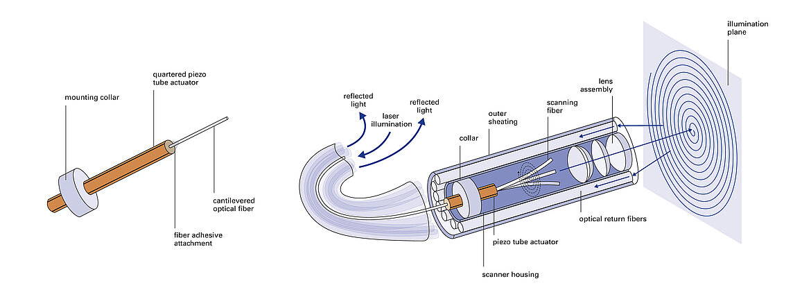
In medicine, finest endoscopes are not only being used for diagnostics but also more and more frequently for the therapy of illnesses. Thanks to their compact and flexible design of just a few millimeters diameter, they ensure even gentler minimally invasive procedures. Contrary to the limitations of the image quality of common endoscopes, the new innovative design of scanning fiber endoscopes (SFEs) offers new possibilities: SFEs generate laser-based, fully colored images with higher resolution and an enlarged visual field, e.g., in optical coherence tomography. The fast motion and control of piezo tubes leads to a scanning motion of the optical fiber in the scanning fiber endoscope. Thanks to this, SFEs deliver more image information, new findings in biomedical research, and improved minimally invasive procedures for the clinical routines.
The central imaging component in scanning fiber endoscopes is a single-mode optical fiber driven by a tubular piezoelectric actuator with segmented electrodes. Miniaturized piezo tubes can realize ultrafast movements using the inverse piezo effect. These segmented piezo tubes not only offer a radial and axial displacement, but a scanning motion in the XY plane can take place with a targeted control of the segments. With that, the optical fiber in the SFE is excited in mechanical resonance between 5 and 12 kHz and the surface to be imaged is scanned with an RGB laser light. The radial vibration movement can be modelled and, subsequently, the optical fiber will move during operation, e.g., in a spiral scan pattern or in Lissajous figures.

The electrical voltages applied during operation of the SFE are low since the tubular piezo actuator electrically corresponds to a small capacitor – in typical operation, the voltage is below 50 V. Similar scanning applications are also possible with bending actuators that achieve relatively large displacements even over the smallest area.
