Adjustment of the Focal Planes in Confocal Microscopy
Content
Adjustment of the Focal Planes in Confocal Microscopy
High-Resolution 3D Imaging
Confocal microscopy is used in live cell imaging to produce virtual 3D sections and Z stacks through individual cells or cell compounds. It is also used in dermatology for in vivo diagnostics of pathological cell alterations close to the skin surface.
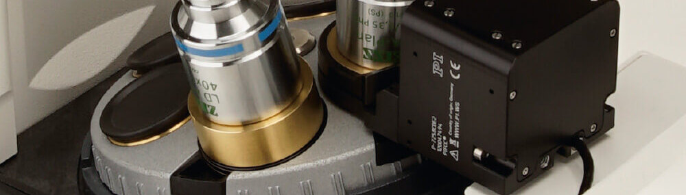
Piezo-Based Drive Solutions
Piezo-based drive solutions position the objective with a repeatability of a few nanometers over ranges of up to several hundred micrometers. The individual images are then superimposed to create a high-resolution 3D image.
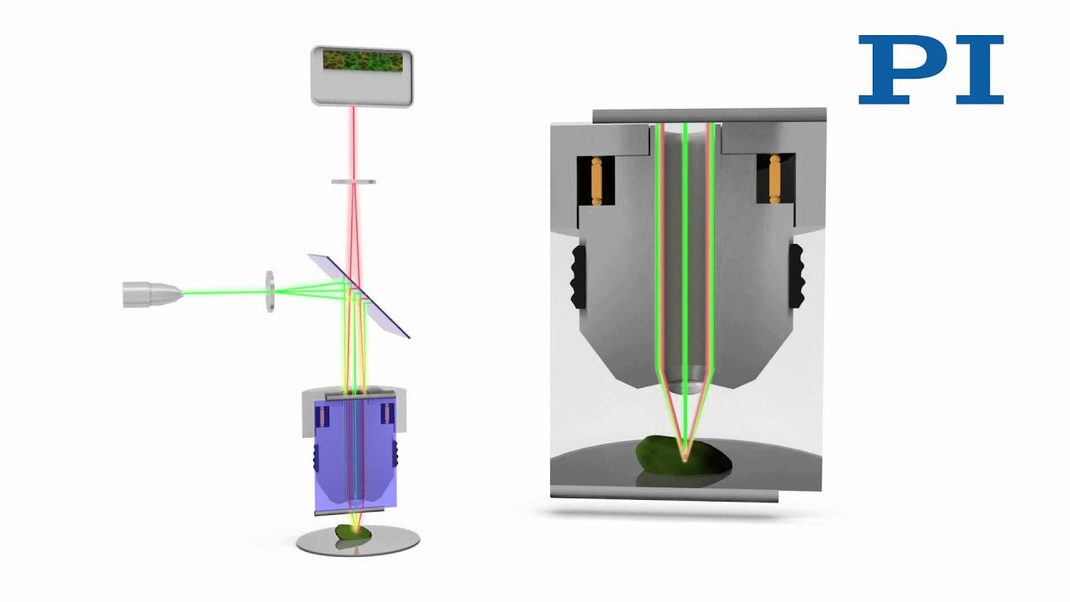
Confocal microscopy is used in diagnostics in ophthalmology and dermatology, for example, to produce virtual sections through the tissue structure, or to detect the structure of the sample surface through the shift of the focal plane. Piezo-based drive solutions position the objective at a distance of less than 1 µm on the different focal planes. The individual images are then superimposed to create a high-resolution 3D image.
Precise Z-positioning for creating Z stacks, 3D images, and sections.
Processes which are observed over time benefit from short settling times in the millisecond range.